

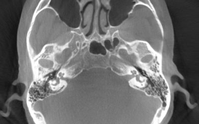
Cervical Spine Imaging
The study (below) is a C-Spine surgical case using Epica’s “SeeFactor CT3”
Sylvan palmer, MD Mission Hospital – Orange County, CA
Spine (Below) Currently, Physicians see this: (MRI)
- MRI is great for seeing and diagnosing nerves, but very poor for bone
- In spine surgery, surgeons are cutting bone, but never see it well
How much could procedural outcomes improve if surgeons could diagnose, plan, and validate intraoperatively using Epica Imaging?

Currently, surgeons see this: spine (intraoperative view)

Surgeons are limited to ‘seeing’ tactilely with instruments and with limited bone information from low-res MRI

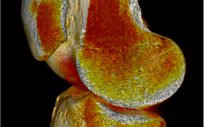
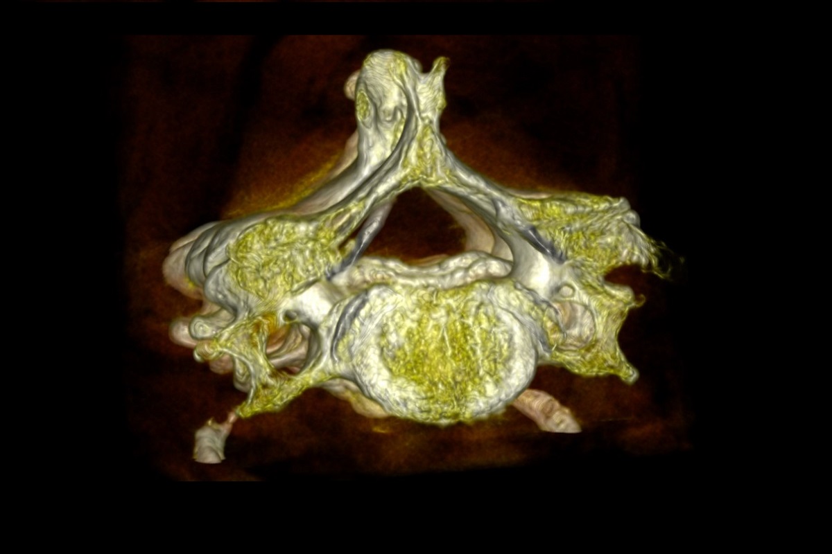
With Epica Imaging, Physicians will see this:
With Epica Imaging, Physicians will have this:
100% Epica Data
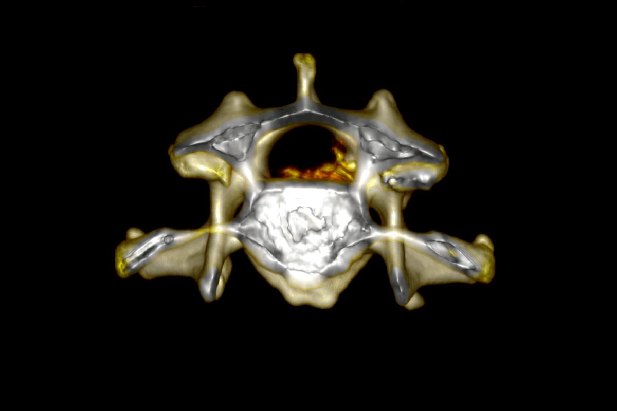
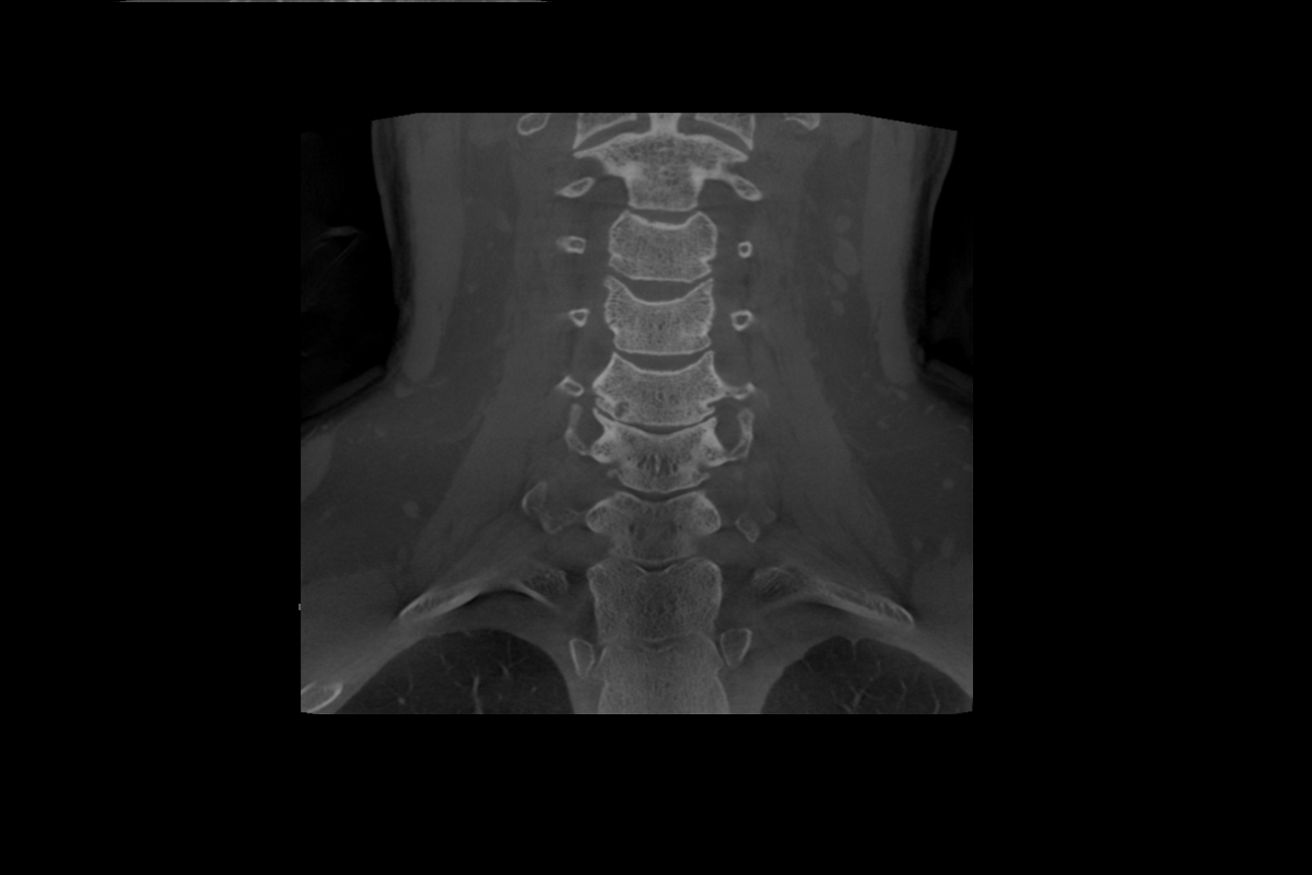
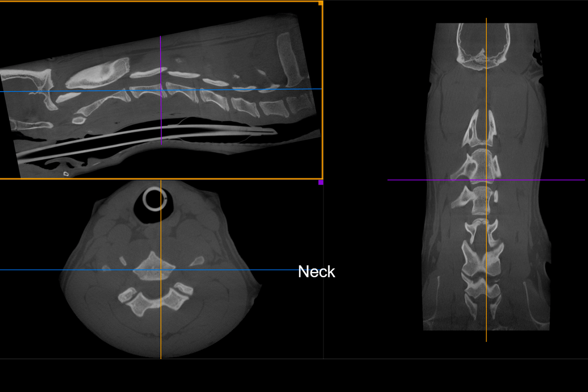
(Below) Frontal C-Spine: Pre-OP Epica SeefactorCT3 Ultra-high Resolution CT Image

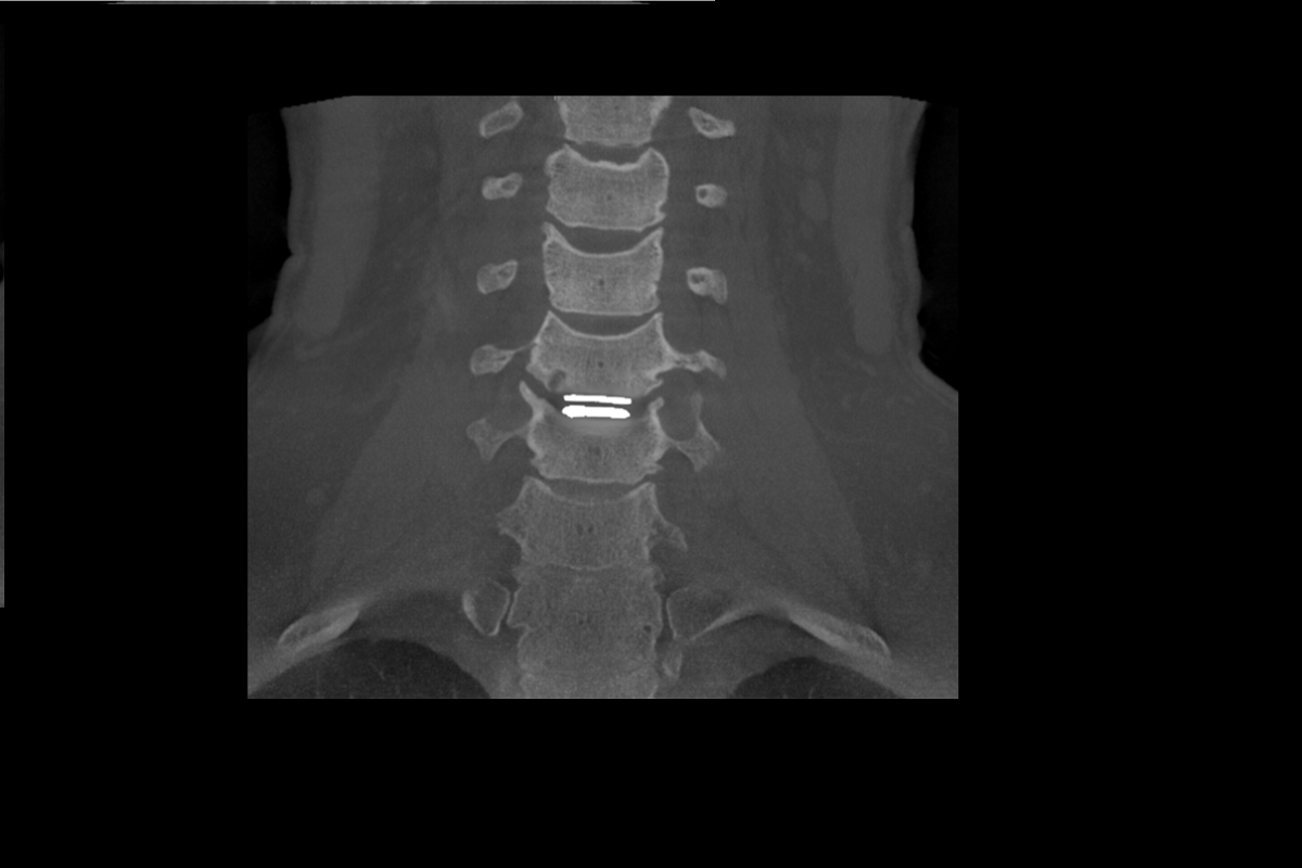
(Below) Frontal C-Spine: Post-OP Epica SeeFactorCT3 Ultra-high Resolution CT Image + Mobi-C Implants

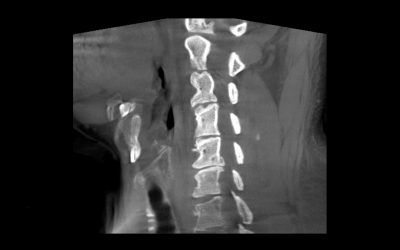
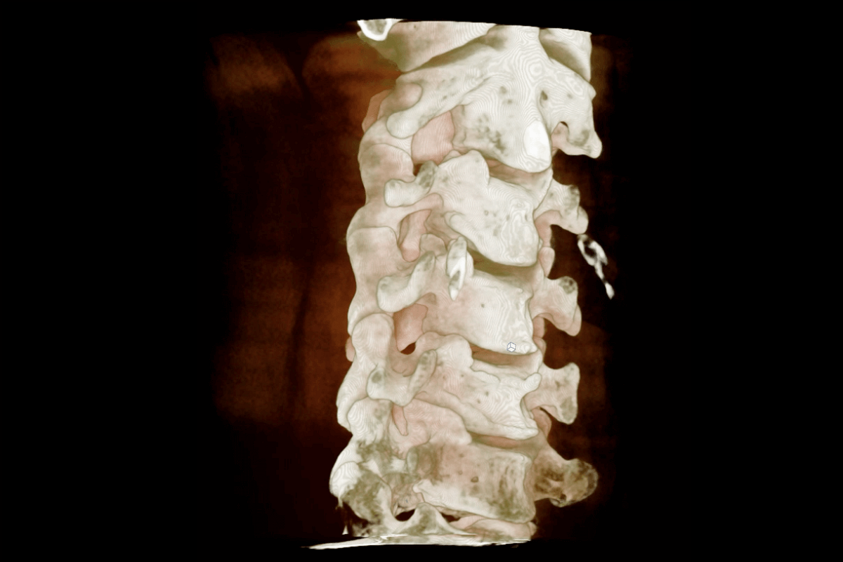
(Below) Sagittal C-Spine: Pre-OP Epica SeeFactorCT3 Ultra-high Resolution CT Image

(Below) Sagittal C-Spine: Post-OP Epica SeeFactorCT3 Ultra-high Resolution CT Image + Mobi-C Implants

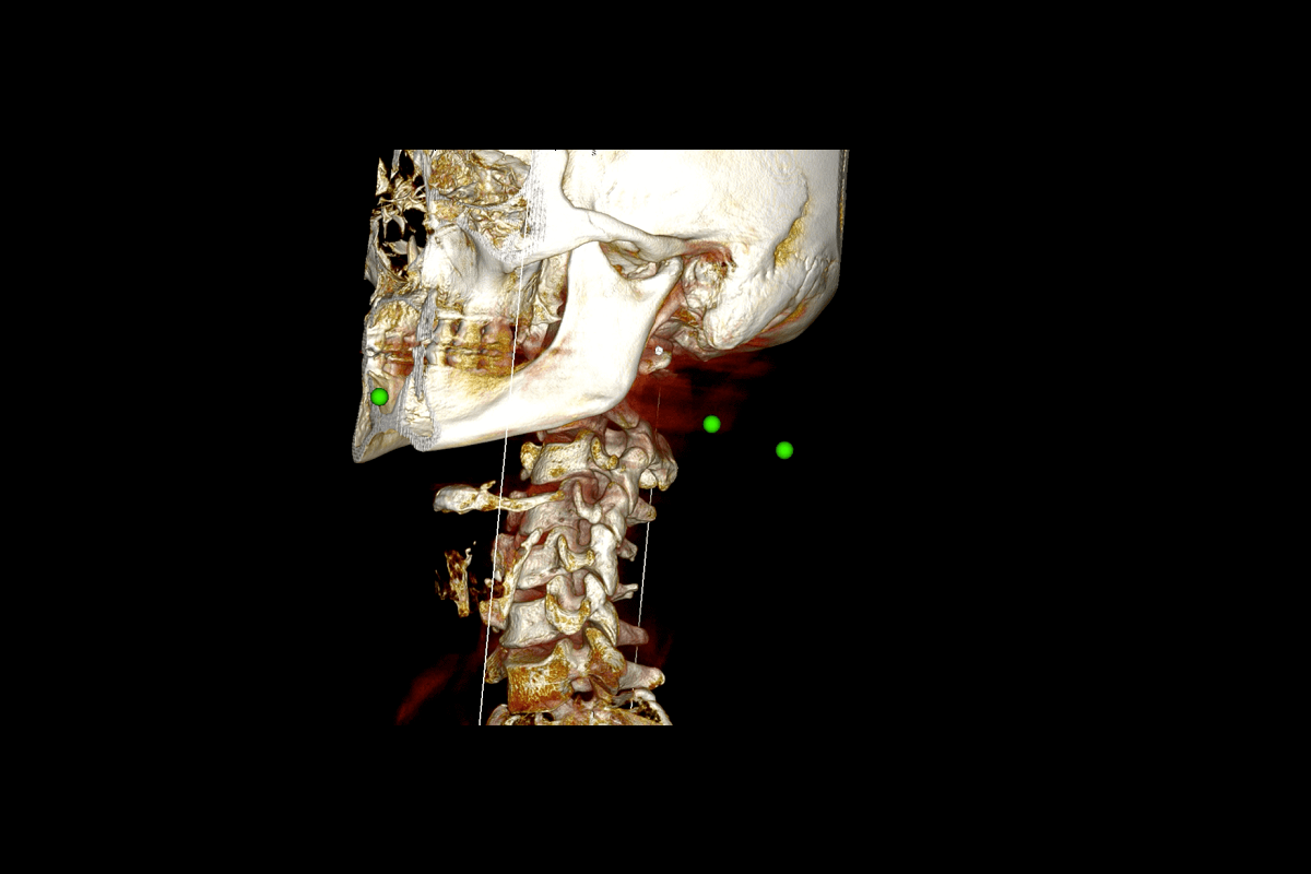
HDVI CT (Volumetric imaging) allows accurate high-resolution measurements to diagnose and validate treatment surgical success. This view has never been seen by physicians, but is critical to successful treatment.
Note: This view is possible only with Epica’s volumetric imaging


0 Comments